Ligaments play a pivotal role in the human body, acting as critical components of the skeletal system. These sturdy bands of fibrous tissue connect bones to other bones at joints, providing essential support and stability. By maintaining the proper alignment and facilitating appropriate movement, ligaments prevent injuries while allowing the flexibility necessary for daily activities and athletic endeavors.
What Are Ligaments?
Ligaments are tough, flexible bands of fibrous connective tissue that connect bones to other bones at joints. They are essential components of the skeletal system, providing stability and support while allowing for necessary movement. Ligaments help to maintain proper alignment and prevent excessive movements that could damage joints and bones.
Types of Ligaments
1. Articular Ligaments
Articular ligaments directly connect bones within a joint, helping to stabilize and guide joint movements. They are typically found surrounding joints such as the knees, elbows, and shoulders. A prime example is the anterior cruciate ligament (ACL) in the knee, which helps control the movement of the joint and provides stability during physical activities.
2. Capsular Ligaments
These ligaments are part of the joint capsule that encloses certain joints. They are thicker parts of the joint capsule and help provide additional stability. Capsular ligaments are crucial in joints that undergo extensive movement, such as the shoulder joint, where they help prevent dislocations and maintain proper joint alignment.
3. Extracapsular Ligaments
Located outside the joint capsule, extracapsular ligaments provide additional support and stability, supplementing the structure of articular and capsular ligaments. They are often found in joints that bear significant weight or endure substantial stress, such as the knee. The lateral collateral ligament (LCL) of the knee is an example, offering stability against varus forces.
4. Intracapsular Ligaments
These ligaments are located inside the joint capsule but are excluded from the joint cavity by the synovial membrane. They are particularly important in the knee joint, where ligaments like the ACL and the posterior cruciate ligament (PCL) play critical roles in stabilizing the joint internally.
5. Elastic Ligaments
Elastic ligaments, such as those found in the vertebral column (ligamenta flava), contain more elastic fibers than typical fibrous ligaments. This composition allows them to stretch considerably and return to their original shape, supporting the body’s flexibility and movement, especially in the spine.
Function of Ligaments
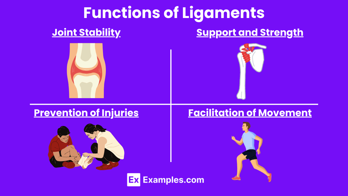
- Joint Stability: Ligaments provide essential stability to joints by preventing excessive or abnormal movements. This helps in maintaining joint integrity and alignment during various activities.
- Support and Strength: By connecting bones, ligaments contribute to the overall strength and support of the skeletal structure, ensuring that the body can withstand physical stresses.
- Facilitation of Movement: Although primarily known for stabilization, ligaments also facilitate proper joint movement. They guide the joints through appropriate ranges of motion, aiding in smooth and coordinated movements.
- Prevention of Injuries: By limiting the range of motion to normal limits, ligaments play a significant role in preventing injuries such as dislocations and sprains.
- Proprioception: Ligaments contain nerve endings that help in proprioception, which is the ability to sense the position and movement of body parts. This sensory feedback helps in coordinating muscle actions and maintaining balance.
Anatomy of Ligaments
Composition and Structure
Ligaments are primarily made up of dense fibrous connective tissue. The main structural components of ligaments include
- Collagen Fibers: These are the predominant element in ligaments, providing strength and flexibility. Type I collagen is most common, offering high tensile strength that resists stretching forces.
- Elastin Fibers: Although less abundant than collagen, elastin fibers allow ligaments to stretch and then return to their original shape. This elasticity is crucial in joints that experience a wide range of motion.
- Ground Substance: This is a gel-like matrix that surrounds the fibers, consisting of proteoglycans and water. It helps in distributing mechanical forces across the ligament to minimize damage.
- Cells: The primary cells in ligaments are fibroblasts, which are responsible for producing and maintaining the extracellular matrix, including collagen and elastin.
Vascular Supply and Nerve Innervation
Ligaments generally have a limited blood supply, which is why they appear white and are known for slow healing processes after injury. Some ligaments, such as the ligamentum flavum, have a richer blood supply, contributing to their elasticity and quicker healing capabilities.
Nerve fibers are also present, which help in proprioception — the body’s ability to perceive its own position in space. These nerve endings help monitor the tension within the ligament and provide feedback to the brain, aiding in the coordination of movement and joint stability.
Attachment and Function
Ligaments attach to the periosteum, a dense layer of vascular connective tissue that covers the bones. This attachment is crucial as it anchors the ligaments firmly to the bones, allowing them to fulfill their role in stabilizing joints. The way ligaments are woven into the periosteum provides a secure attachment that supports the joint under various physical stresses.
Functional Adaptability
Ligaments can adapt to changes in the mechanical demands placed upon them. Regular physical activity can lead to a strengthening of these structures, whereas a lack of movement can lead to their weakening. This adaptability highlights the importance of regular exercise in maintaining joint stability and ligament health.
Examples of Ligaments
1.Anterior Cruciate Ligament (ACL)
- Location: Knee joint
- Function: Stabilizes the knee by preventing the tibia (shinbone) from sliding out in front of the femur (thighbone).
2. Posterior Cruciate Ligament (PCL)
- Location: Knee joint
- Function: Works with the ACL to prevent the tibia from sliding backwards under the femur.
3. Lateral Collateral Ligament (LCL)
- Location: Outside of the knee
- Function: Provides lateral stability to the knee, preventing it from bending outward.
4. Medial Collateral Ligament (MCL)
- Location: Inside of the knee
- Function: Provides stability to the inner knee, preventing it from bending inward.
5. Rotator Cuff Ligaments
- Location: Shoulder joint
- Function: Stabilizes the shoulder and holds the head of the humerus in the glenoid cavity of the scapula.
6. Ligamentum Flavum
- Location: Along the spine between vertebrae
- Function: Connects the laminae of the vertebrae and preserves the upright posture, assisting with the flexibility and protection of the spinal column.
7. Calcaneofibular Ligament
- Location: Ankle
- Function: Connects the fibula (smaller bone of the lower leg) to the calcaneus (heel bone) and stabilizes the ankle joint.
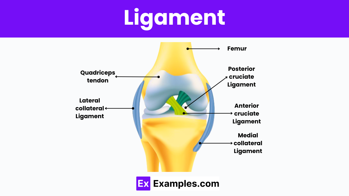
Ligaments of the Knee
The knee joint is stabilized by several crucial ligaments that include the Anterior Cruciate Ligament (ACL), Posterior Cruciate Ligament (PCL), Medial Collateral Ligament (MCL), and Lateral Collateral Ligament (LCL). The ACL and PCL cross each other inside the knee, working together to control the forward and backward motion of the tibia (shinbone) relative to the femur (thighbone). The MCL, located on the inner side of the knee, and the LCL, on the outer side, manage the sideways movements and overall stability of the knee. These ligaments are essential for maintaining knee stability during movement, supporting dynamic activities like walking, running, and turning, and protecting the knee from unnatural movements that could lead to injury.
Ligament Injury and Tear
Ligament injuries are common, particularly among athletes and individuals engaged in physical activities, but they can also occur during everyday activities. These injuries can range from mild sprains to severe tears, affecting the stability and function of the affected joint. Ligament injuries typically occur when a joint is stressed beyond its normal range of motion, often due to sudden twisting, stretching, or impact. Here’s how these injuries are classified
- Sprains: These are injuries to ligaments caused by being stretched past their normal capacity. Sprains are graded based on their severity:
- Grade I (mild): Slight stretching and microscopic tearing of the ligament fibers. There is minimal tenderness and swelling.
- Grade II (moderate): Partial tearing of the ligament. This can cause noticeable swelling, bruising, and tenderness at the joint, along with a loose feeling when moving it.
- Grade III (severe): Complete tear of the ligament leading to joint instability. This grade often makes the joint non-functional.
Ligaments of the Human Body
- Anterior Cruciate Ligament (ACL) and Posterior Cruciate Ligament (PCL): Located in the knee, these ligaments are key to stabilizing the joint. The ACL prevents the tibia from sliding too far forward, and the PCL stops it from sliding too far backward.
- Medial Collateral Ligament (MCL) and Lateral Collateral Ligament (LCL): Also part of the knee, the MCL and LCL prevent the leg from overextending inward or outward, respectively.
- Rotator Cuff Ligaments: These include a group of ligaments that surround the shoulder joint, securing the head of the humerus within the shallow socket of the shoulder.
- Ankle Ligaments: Including the anterior talofibular ligament (ATFL), posterior talofibular ligament (PTFL), and calcaneofibular ligament (CFL), these support the ankle joint and help in its range of motion.
Artificial Ligaments
Artificial ligaments are synthetic structures designed to mimic the function of natural ligaments in the human body. These are typically used in cases where natural ligament repair is not possible or where previous surgeries have failed. Artificial ligaments offer an alternative to traditional surgical treatments like tendon grafts and are an area of significant interest in orthopedic and sports medicine.
Development and Materials
The development of artificial ligaments has evolved over the years with advances in materials science and biomedical engineering. These synthetic ligaments are usually made from biocompatible materials that can withstand the mechanical stresses placed on joints during movement. Common materials include:
- Polyethylene terephthalate (PET): Often used for its durability and strength.
- Polypropylene: Known for its elasticity and fatigue resistance.
- Polytetrafluoroethylene (PTFE): Also known as Teflon, used for its low friction properties.
- Carbon fibers: Praised for their high stiffness and tensile strength.
FAQs
What is the Difference Between a Tendon and a Ligament?
Tendons connect muscles to bones, while ligaments connect bones to bones, providing joint stability.
What Happens When a Ligament is Broken?
A broken ligament, often called a tear, causes joint pain, swelling, and instability.
Can a Ligament Heal on Its Own?
Minor ligament injuries can heal on their own with rest and proper care, but severe tears may require surgery.
How Long Does It Take Ligaments to Heal?
Ligament healing time varies, generally taking several weeks to months depending on injury severity and treatment.
What is the Fastest Way to Heal Ligaments?
Prompt medical treatment, rest, ice, compression, elevation (RICE), and physical therapy are the fastest ways to heal ligaments.