
Cilia are microscopic, hair-like structures that extend from the surface of many types of cells in the bodies of various organisms, including humans. These tiny organelles play a crucial role in cellular functions, from movement to sensory processes. Cilia can be classified into two main types: motile cilia, which are primarily involved in movement either of the cells themselves or of fluids surrounding the cells, and non-motile or primary cilia, which serve as sensory organelles. This article delves into the structure, function, and significance of cilia, highlighting their essential contributions to health and disease management. By understanding cilia, we gain insights into their critical roles across a wide range of biological processes and systems.
What is Cilia?
Cilia are slender, microscopic structures that resemble tiny hairs, which protrude from the surface of certain cells. They are a fundamental component of many living organisms, including humans. Structurally, each cilium is composed of a core called the axoneme, which consists of a set of microtubules arranged in a specific pattern, typically a “9+2” structure. This pattern includes nine doublet microtubules encircling two central singlet microtubules.
Structure of Cilia
1. Axoneme
The core structural component of a cilium is the axoneme, which serves as the backbone. The axoneme typically follows a “9+2” arrangement consisting of nine pairs of microtubules (doublets) arranged around a central pair of single microtubules (singlets). This arrangement is crucial for the movement capabilities of motile cilia through the coordinated sliding of the microtubules, driven by the motor protein dynein.
2. Basal Body
The axoneme of the cilium originates from the basal body, which is derived from a centriole, a key component of the cell’s cytoskeleton. The basal body anchors the cilium to the cell and ensures its structural stability. It acts as a foundation, organizing the growth and maintenance of the axoneme.
3. Transition Zone
Situated between the basal body and the axoneme, the transition zone functions as a gateway that regulates the movement of molecules in and out of the cilium. This zone is critical for maintaining the chemical environment within the cilium, which is necessary for its sensory and movement functions.
4. Ciliary Membrane
Encasing the axoneme is the ciliary membrane, which is continuous with the plasma membrane of the cell but has a unique composition of lipids and proteins. This specialized membrane plays a key role in signal transduction for non-motile cilia and in protecting the structural integrity of the cilium.
5. Motor Proteins and Nexin Linkers
Motor proteins, such as dynein, are essential for the movement of motile cilia. Dynein arms attached to one microtubule can grab onto the adjacent microtubule and walk along it, causing the microtubules to slide relative to each other, which results in the bending of the cilium. Nexin linkers serve as bridging molecules that connect adjacent doublet microtubules, providing elasticity and preventing them from sliding too far apart.
6. Y-Links and Radial Spokes
These structures provide additional support and regulation within the cilium. Y-links connect the outer doublet microtubules to the ciliary membrane, helping maintain the structural integrity of the cilium. Radial spokes extend from the microtubules to the central sheath surrounding the central pair, playing a role in regulating the movement and stiffness of the axoneme.
Types of Cilia
1. Motile Cilia
Motile cilia are primarily responsible for generating movement. They are usually found in large numbers on the surface of cells and move in a coordinated, wave-like fashion. Here are key features and functions of motile cilia:
- Location and Function: These cilia line the respiratory tract where they help sweep mucus and trapped particles out of the lungs, aiding in respiratory health. In the reproductive system, motile cilia help transport the egg from the ovary through the fallopian tube towards the uterus.
- Structure: Motile cilia contain the “9+2” microtubule arrangement typical of ciliary axonemes, which is essential for their ability to beat rhythmically.
2. Non-Motile (Primary) Cilia
Non-motile cilia, also known as primary cilia, typically occur singly on most cells and do not participate in generating movement. Instead, they serve crucial sensory and signaling functions. Characteristics include:
- Sensory Capabilities: These cilia are involved in signal transduction, acting as the cell’s antenna for sensing the external environment. They are crucial in processes ranging from growth control to cell division and differentiation.
- Structure: Unlike motile cilia, primary cilia usually feature a “9+0” arrangement, lacking the central pair of microtubules found in motile cilia. This structure is adapted more for stability and sensory functions rather than movement.
- Health Implications: Defects in primary cilia can lead to various genetic disorders known as ciliopathies, which include conditions like polycystic kidney disease and Bardet-Biedl syndrome.
3. Specialized Cilia
In addition to these main types, some specialized forms of cilia exist, tailored to specific functions:
- Nodal Cilia: These are found in the embryonic node of vertebrates and play a role in left-right asymmetry determination in developing embryos. Nodal cilia are motile but have a unique rotational movement rather than the usual wave-like motion of typical motile cilia.
Functions of Cilia
Movement and Locomotion
One of the primary functions of cilia is to facilitate movement. In single-celled organisms like Paramecium, cilia cover the cell’s surface, beating rhythmically to propel the organism through water. In humans, cilia are found in the respiratory tract, where they execute a coordinated waving motion to transport mucus and trapped particles out of the airways, helping to clear inhaled pathogens and debris.
Sensory Roles
Cilia are not just mechanical performers; they also serve as sensory organelles. For example, they are present in the kidney where they detect fluid flow through the renal tubules, contributing to signaling pathways that regulate kidney function. In the eye, specifically in the retina, modified cilia play a role in the structure and function of photoreceptor cells, which are crucial for vision.
Developmental Signaling
During development, cilia are involved in several signaling pathways that are crucial for the proper growth and organization of the body. The Hedgehog signaling pathway, essential for embryonic development, relies on the presence of cilia to function properly. Defects in ciliary structure or function can lead to developmental disorders such as polycystic kidney disease and congenital heart defects.
Fluid Circulation
In certain organs, cilia contribute to the circulation of fluids. For example, in the brain, cilia help move cerebrospinal fluid, which is crucial for cushioning the brain and removing waste products. In the female reproductive system, cilia within the fallopian tubes move the egg from the ovary towards the uterus.
Health Implications
Given their extensive roles, it’s evident that cilia are vital for maintaining normal physiological functions. Dysfunction of cilia, known as ciliopathies, can lead to a wide array of health issues, including respiratory problems, kidney disease, and fertility issues. Understanding the function of cilia can aid in diagnosing and treating these conditions more effectively.
Cilia in Lungs
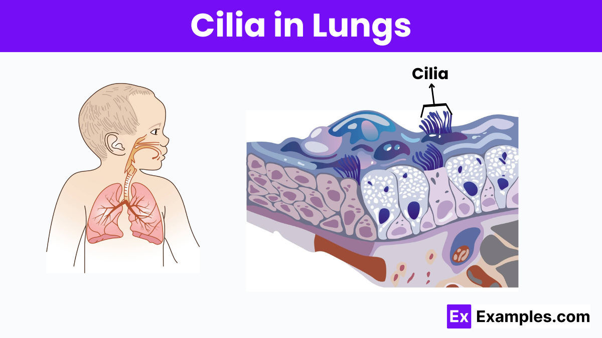
Cilia in the lungs play a crucial role in respiratory health, acting as tiny, hair-like structures that line the airways. Their main function is to trap and move particles and pathogens out of the airways, helping to prevent infections and maintain clear air passages. This movement, often compared to a wave, efficiently propels mucus loaded with dust, bacteria, and other foreign particles upward toward the throat, where it can be swallowed or expelled. The proper functioning of cilia is essential for protecting the lungs from irritants and infections, highlighting their importance in respiratory defense mechanisms.
Cilia in the Human Body
Cilia are microscopic, hair-like structures that extend from the surface of many types of cells in the human body.
Movement
In the respiratory system, cilia help move mucus and trapped particles out of the airways, aiding in the prevention of respiratory infections by clearing debris and pathogens.
Sensory Functions
Cilia in the kidney cells detect fluid flow, which is essential for regulating kidney function and maintaining a balance of body fluids.
Developmental Processes
Cilia are involved in important signaling pathways during embryonic development, influencing the proper growth and organization of tissues and organs.
Fluid Circulation
Cilia in the brain help move cerebrospinal fluid, playing a role in protecting brain tissues and removing waste.
Ciliogenesis – Formation of Cilia
Ciliogenesis is the biological process responsible for the formation of cilia on the surface of cells. Cilia are tiny, hair-like structures that protrude from the cell membrane and are found in almost all mammalian cells. They play crucial roles in various cellular functions, including motility, sensory perception, and signal transduction.
The Stages of Ciliogenesis
1. Initiation of Ciliary Vesicle Formation: The process of ciliogenesis begins with the formation of a ciliary vesicle, which originates from the cell’s centrosome. The centrosome, acting as the main microtubule organizing center, anchors and stabilizes the base of the emerging cilium.
2. Migration of the Basal Body: Following vesicle formation, the mother centriole of the centrosome migrates to the cell surface. This centriole transforms into the basal body, which acts as a template for cilia construction. The migration is facilitated by various motor proteins and microtubule dynamics.
3. Axoneme Extension: The axoneme, the central shaft of the cilium, begins to extend from the basal body. It is composed of microtubule doublets arranged in a specific pattern, usually a “9+2” arrangement in motile cilia and a “9+0” in primary cilia. The extension of the axoneme is powered by the addition of tubulin proteins, which elongate the microtubules.
4. Ciliary Membrane Formation: As the axoneme extends, the ciliary membrane grows around it, separating the cilium from the cytoplasm of the cell. The membrane is sourced from the cell’s own plasma membrane but develops a distinct composition of proteins and lipids.
5. Maturation and Differentiation: The final stage of ciliogenesis involves the maturation of the cilium and its differentiation into either a motile or sensory cilium. Motile cilia acquire additional structures like radial spokes and dynein arms, necessary for their movement. Sensory cilia, on the other hand, develop receptor proteins and signaling complexes essential for cellular communication and environmental sensing.
Clinical Significance of Cilia
Role in Respiratory Health
In the respiratory system, cilia are crucial for clearing mucus and foreign particles. This mucociliary clearance is vital for protecting the lungs from infections and maintaining clear airways. Dysfunction in ciliary activity, such as in primary ciliary dyskinesia (PCD), leads to recurrent respiratory infections, sinusitis, and bronchiectasis.
Impact on Reproductive Health
Cilia are also essential in the reproductive systems. In females, cilia within the fallopian tubes facilitate the movement of the egg from the ovary to the uterus. Impaired ciliary function can result in ectopic pregnancies or infertility. Similarly, in males, cilia affect sperm motility and can influence fertility outcomes.
Influence on Developmental Processes
Cilia play a significant role in developmental signaling pathways. For example, cells modulate the sonic hedgehog (SHH) signaling pathway, crucial for normal organ development, through their cilia. Mutations that affect ciliary structure or function can lead to developmental disorders such as holoprosencephaly, where the brain does not divide properly into two hemispheres.
Association with Kidney Function
In the kidneys, cilia sense fluid flow within nephrons, the functional units of the kidney. This sensory function is crucial for normal kidney development and function. Defects in ciliary function are linked to polycystic kidney disease (PKD), which is characterized by the growth of numerous cysts that can lead to chronic kidney disease.
Connection to Neurological Functions
In the brain, particularly on neurons, cilia are implicated in neurological functions and behaviors. Abnormalities in these cilia can associate with conditions such as Bardet-Biedl syndrome and Joubert syndrome, which present a range of neurological symptoms, including developmental delays and coordination problems.
Cilia Disorders
Cilia disorders, commonly known as ciliopathies, arise from structural or functional abnormalities in cilia. These microscopic, hair-like structures are essential for various cellular functions, including motility, fluid movement, and signaling pathways. Ciliopathies can affect multiple organ systems, leading to a wide range of symptoms and health issues.
Primary Ciliary Dyskinesia (PCD)
Primary Ciliary Dyskinesia is a genetic disorder characterized by chronic respiratory tract infections, sinusitis, and reduced fertility. Dysfunctional motile cilia fail to clear mucus and bacteria from the respiratory tract effectively.
Polycystic Kidney Disease (PKD)
Polycystic Kidney Disease involves the growth of numerous cysts in the kidneys, impairing their function. This condition links to defects in primary cilia, which normally help in sensing fluid flow and maintaining proper kidney architecture and function.
Bardet-Biedl Syndrome
Bardet-Biedl Syndrome is a ciliopathy with symptoms including obesity, pigmentary retinopathy, polydactyly, renal abnormalities, and learning difficulties. Mutations affecting cilia’s sensory and signaling roles cause it.
Joubert Syndrome
Joubert Syndrome is marked by a distinctive cerebellar malformation visible on MRI scans, known as the “molar tooth sign.” Affected individuals may exhibit poor muscle tone, coordination difficulties, and developmental delays, reflecting the importance of cilia in neural development.
Meckel-Gruber Syndrome
Meckel-Gruber Syndrome is a severe ciliopathy characterized by kidney cysts, liver fibrosis, and neurological abnormalities. It is typically fatal, demonstrating the critical role cilia play in organ development and function.
FAQs
What is Cilia and Its Function?
Cilia are microscopic, hair-like structures on cell surfaces that move fluids or sense environmental signals, crucial for cellular function and health.
What Are Cilia and Why Are They Important?
Cilia are essential cell structures that filter air, remove contaminants, and are vital for sensory functions, impacting overall health and disease prevention.
What Two Things Are Cilia Used For?
Cilia are used for locomotion, helping cells move, and for sensing, where they detect changes in the cell’s environment.
What Are Cilia in the Lungs?
In the lungs, cilia sweep away mucus and pathogens, protecting respiratory passages from infection and buildup.
Do Lung Cilia Grow Back?
Yes, damaged lung cilia can regenerate and restore function, ensuring continued protection against inhaled pathogens and debris.